File:Rod.jpg
| Uploaded by | Nuvitauy07 |
|---|---|
| Upload date | 2009-08-02T21:21:43Z |
| MIME type | image/jpeg |
| Dimensions | 326 × 382 px |
| File size | 32.6 KB |
Summary
| Description |
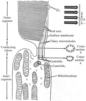
English: Diagram of a mammalian retinal rod cell, showing the centriole pair associated with the modified cilium of the outer segment. The outer segment contains rod sacs that are transversely arranged membranous disks formed from the folded ciliary membrane. |
| Date | September 2000, Updated July 2006, Updated November 2008 |
| Source | http://light.simanonok.com/RetinalRod.jpg |
| Author | Avers, 1976, p 387 |

Licensing


This file is licensed under the Creative Commons Attribution-Share Alike 3.0 Unported license.
- You are free:
- to share – to copy, distribute and transmit the work
- to remix – to adapt the work
- Under the following conditions:
- attribution – You must give appropriate credit, provide a link to the license, and indicate if changes were made. You may do so in any reasonable manner, but not in any way that suggests the licensor endorses you or your use.
- share alike – If you remix, transform, or build upon the material, you must distribute your contributions under the same or compatible license as the original.